


Lumbar osteochondrosis is a chronic disease that develops as a result of the degenerative-dystrophic process of the intervertebral discs. The disease is widespread and affects most people between the ages of 25 and 40.
Statistics show that every second adult experiences back pain at least once in their lifetime, while in 95% of cases it causes osteochondrosis of the spine.
Patients with severe lumbar osteochondrosis, persistent pain, and other manifestations are temporarily disabled. If their condition does not improve within four months, the question of creating a disability group will be decided.
Lumbar osteochondrosis is a serious medical and social problem, as the disease primarily affects most people of working age, and in the absence of treatment, it can cause the development of a disc herniation.
Causes and risk factors
Predisposing factors for lumbar osteochondrosis:
- anomalies in the structure of the spine;
- lumbarization - a congenital pathology of the spine characterized by separation of the first vertebra from the sacrum and its transformation into the sixth (additional) lumbar region;
- sacralization is an innate pathology in which the fifth lumbar vertebrae merges with the sacrum;
- asymmetrical arrangement of the joint spaces of the intervertebral joints;
- abnormal narrowing of the spinal canal;
- reflects spondiogenic pain (somatic and muscle);
- obesity;
- sedentary lifestyle;
- prolonged vibration;
- systematic physical stress;
- smoking.
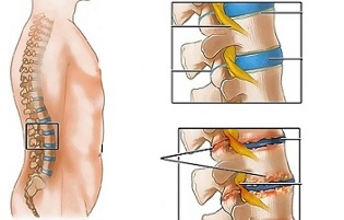
Adverse static-dynamic loads, combined with one or more risk factors, alter the physiological properties of the pulp of the fibrous disc core, which plays a shock-absorbing role and ensures spinal mobility. This process is based on the depolymerization of polysaccharides, which leads to loss of moisture in the gelatinous tissue. As a result, the nucleus pulposus and with it the fibrous disc lose its elasticity. Additional mechanical loads provoke elongated ring fibrosis protrusion. This phenomenon is called protrusion. Cracks appear in the fibrous nucleus through which fragments of the pulposous nucleus fall out (prolapse, herniated plate).
Prolonged compression of nerve roots, which over time innervates certain abdominal organs, leads to deterioration in their function.
Spinal instability is accompanied by reactive changes in the body of adjacent vertebrae, the intervertebral joints, and with it the development of spondyloarthrosis. For example, significant muscle contraction in the background of physical activity leads to displacement of vertebral bodies and entrapment of nerve roots with the development of radical syndrome.
Another cause of pain and neurological symptoms in lumbar osteochondrosis may be osteophytes - bone outgrowths in processes and vertebral bodies that cause radical syndrome or compression myelopathy (spinal cord compression).
Forms of the disease
Depending on which structures are involved in the pathological process, lumbar osteochondrosis is clinically manifested in the following syndromes:
- reflex- lumbodynia, lumboishalgia, lumbago; they develop in the background of reflex overload of the back muscles;
- compression (spine, blood vessel, radical)- their development is caused by compression (compression) of the spinal cord, blood vessels or nerve roots. Examples are lumbosacral radiculitis and radiculoischemia.
Symptoms of lumbar osteochondrosis
In lumbar osteochondrosis, symptoms are determined by which structures are involved in the pathological process.
Lumbago occurs as a result of hypothermia or physical overload, and sometimes for no reason. The pain suddenly appears and is shooting in nature. It increases when sneezing, coughing, turning the body, exercising, sitting, standing, walking. In the supine position, the pain sensation was significantly weakened. Sensitivity and reflexes are maintained, and the range of motion of the lumbar spine decreases.
Observe the feel:
- pain in the lumbar region;
- spasm of the paravertebral muscles;
- flattening of lumbar lordosis, which in many cases is combined with scoliosis.
Nerve root tension syndrome is negative. When the straight leg is lifted, patients experience an increase in pain in the lumbar region rather than their appearance in the outstretched lower limb.
In lumbar osteochondrosis, pain attacks often recur, each time becoming more intense and prolonged.
For lumbodynia, the clinical picture is similar to lumbago, but the increase in pain intensity occurs over several days.
In lumboishalgia, patients complain of radiating pain to one or both lower extremities of the lumbar region. The pain spreads to the buttocks and back of the thighs and never reaches the legs.
Lumboishalgia is characterized by vasomotor disorders:
- changes in lower limb skin temperature and color;
- feeling warm or cool;
- violation of blood circulation.
The development of lumbar compression syndromes is clinically indicated by the following symptoms:
- dermatological hypalgesia;
- shot pains;
- weakening or complete loss of deep reflexes;
- peripheral paresis.
In compression syndromes, pain is exacerbated by trunk bending, sneezing, and coughing.
Diagnostics
Diagnosis of lumbar osteochondrosis is based on data from the clinical picture of the disease, laboratory and instrumental research methods.
In blood tests for lumbar osteochondrosis:
- decrease in calcium concentration;
- increased ESR;
- increased alkaline phosphatase levels.
X-ray examination of the spine is of great importance in the diagnosis of lumbar osteochondrosis.
Prolonged compression of nerve roots, which over time innervates certain abdominal organs, leads to deterioration in their function.
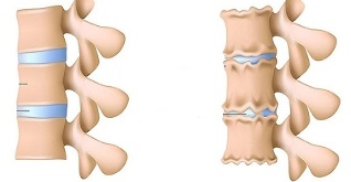
The X-rays that confirm the diagnosis are as follows:
- change the configuration of the affected segment;
- pseudospondylolisthesis (displacement of adjacent vertebral bodies); deformation of the closing plates
- ;
- smoothing the intervertebral disc;
- is the unequal height of the intervertebral disc (a symptom of spacer) associated with asymmetric muscle tone.
It is also used in the diagnosis of lumbar osteochondrosis if it is indicated:
- myelography, computer or magnetic resonance imaging - necessary for persistent symptoms, neurological deficits;
- scintigraphy (examination of the accumulation of phosphorus in the skeletal system, marked tech-99) - occurs when a tumor or infectious process, spinal cord injury is suspected.
Differential diagnosis of lumbar osteochondrosis is made with the following diseases:
- spondylolisthesis;
- dyshormonal spondylopathy;
- ankylosing spondylitis (ankylosing spondylitis);
- infectious processes (disc inflammation, spinal osteomyelitis);
- tumor processes (primary tumor of the spine or metastatic lesions);
- rheumatoid arthritis;
- deforming hip joint osteoarthritis;
- reflected pain (diseases of the internal organs and large blood vessels).
Treatment of lumbar osteochondrosis
In lumbar osteochondrosis, the following tactics are usually followed:
- bed rest for 2-3 days; dragging the affected segment of the spine
- ;
- strengthening the back and abdominal muscles (creating a so-called muscle ligament);
- effect on abnormal myofascial and myotonic processes.
Lumbago occurs as a result of hypothermia or physical overload, and sometimes for no reason.
In most cases, conservative treatment of lumbar osteochondrosis is performed, including the following measures:
- muscle infiltration anesthesia with a solution of local anesthetics;
- taking non-steroidal anti-inflammatory drugs; taking
- sensitizers;
- vitamin therapy;
- taking sedatives and antidepressants;
- manual therapy, massage;
- physiotherapy exercises;
- acupuncture;
- post-isometric relaxation.
The absolute indications for surgical treatment of lumbar osteochondrosis are:
- acute or subacute spinal cord compression;
- Development of cauda equina syndrome characterized by dysfunction of pelvic organs, sensory and motor disorders.
Therapeutic practices for lumbar osteochondrosis
Physiotherapy plays a significant role in the treatment of lumbar osteochondrosis complex. Regular exercises make it possible to normalize the muscle tone of the paravertebral muscles, improve the metabolic processes in the tissues affected by the pathological process, and also form a well-developed muscle ligament that can support the spine in the right position, placing unnecessary static loads on it.
In order for lumbar osteochondrosis exercise to have the greatest effect, the following principles should be followed:
regularity of- lessons;
- gradual increase in physical activity;
- Avoid class overload.
Physiotherapy should be performed under the guidance of an experienced instructor who selects the exercises that are most effective for the patient and verifies that they are performed correctly.
Statistics show that every second adult experiences back pain at least once in their lifetime, while in 95% of cases it causes osteochondrosis of the spine.
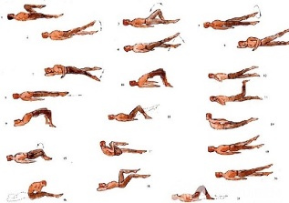
In addition to the lessons with the instructor, perform a series of morning exercises each day that include special exercises for lumbar osteochondrosis.
- Relaxation and contraction of the abdominal muscles.The starting position is, the legs are shoulder-width apart, the arms are lowered to the body. Take a smooth breath, relaxing the muscles of the anterior abdominal wall. During exhalation, tighten your stomach as much as possible, straining your abdominal muscles. The exercise should be repeated until mild fatigue appears.
- Head movements with spine flexion.The starting position is on your knees with your arms outstretched and your back straight. Slowly raise your head and bend your back. Hold this position for a few seconds, then smoothly return to the starting position. Repeat at least 10-12 times.
- "Pendulum".Starting position lying on your back, arms along the body, legs bent at right angles to the knees and hips. Turn your legs to the right and left with swinging pendulum movements, trying to reach the floor. In this case, the shoulder blades cannot be torn off the floor.
- Ship.Starting position lying on your stomach, arms outstretched. Tear off the upper body and legs from the floor, bent at the back. Hold this position for 5-6 seconds and slowly return to the starting position. Run 10 times.
Possible Consequences and Complications
The main complications of lumbar osteochondrosis are:
- intervertebral hernia formation;
- vegetative-vascular dystonia; spondylolysis, spondylolisthesis;
- osteophytosis;
- spondyloarthrosis; Narrowing of the spinal canal
- , which results in compression of the spinal cord, can cause permanent disability and decreased quality of life.
Prolonged compression of nerve roots, which over time innervates certain abdominal organs, leads to a deterioration in their function. As a result, patients have intestinal disorders (constipation, diarrhea, bloating) and pelvic organs (urinary disorders, erectile dysfunction, frigidity, infertility).
Forecast
Pain syndrome in lumbar osteochondrosis occurs in the form of remissions and exacerbations. Lumbago lasts for 10-15 days, after which the patient's condition improves and the pain subsides. A favorable result may be prevented by associated secondary diseases. Often, in the case of lumbar osteochondrosis, the pain attacks recur, becoming more intense and protracted each time.
Physiotherapy plays a significant role in the treatment of lumbar osteochondrosis complex.
Patients with severe lumbar osteochondrosis, persistent pain, and other manifestations are temporarily disabled. If their condition does not improve within four months, the question of creating a disability group will be decided.
Prevention
Prevention of spinal osteochondrosis consists of the following measures:
- quit smoking;
- weight normalization;
- improving general physical condition, active lifestyle;
- Avoid provocative conditions (weightlifting, sudden movements, twists, turns).